


Cell Painting for Morphological Profiling
Unlock the full phenotypic signature of your compounds and targets
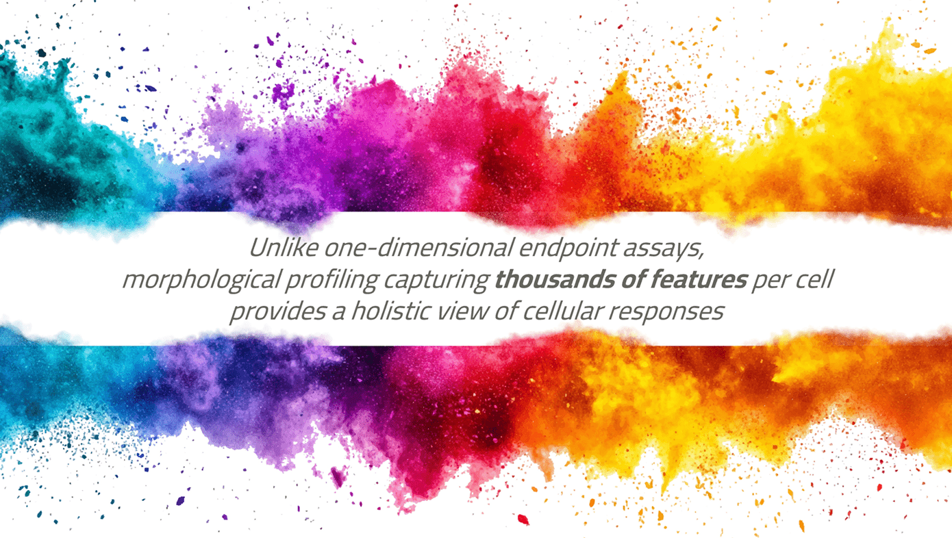
Cell painting is an unbiased, multiplexed, image-based profiling method that simultaneously “paints” multiple cellular structures. It combines automated microscopy with advanced image analysis to quantify subtle changes in morphology, texture, organelle structure, and spatial relationships. This “phenotypic barcode” enables comparison of compounds, inference of biological pathways, toxicity detection, and more.
Why and when use cell painting in drug discovery
Cell painting provides deep phenotypic insights that go beyond traditional single-readout assays. It is ideal for:
- Comprehensive insights beyond single readouts: it captures holistic cell state, not just one biomarker
- Target identification: to identify targets by matching observed phenotype to known target profiles
- Early toxicity detection: phenotypic perturbations often precede viability loss
- Mechanism of action inference via similarity: compare test compounds with reference libraries
- Scalability throughput & relevance: our platform supports moderate to high throughput (e.g. 96/384) with several type of cell models, from recombinant to iPS-derived cells
- Compatibility with AI / ML pipelines: rich high-dimensional data yields powerful models

UMAP clustering and representative cell image overlays. UMAP map of cellular phenotypes, where each dot reflects compound-induced morphological changes. Highlighted groups reveal distinct effects such as cytoskeletal remodeling and mitochondrial stress.
Axxam cell painting capabilities
At Axxam, we offer a comprehensive and flexible cell painting platform tailored to your research needs:
- Canonical 6-channel palettes, in accordance with the JUMP consortium protocol v3: nucleus (Hoechst), ER (ConA), RNA (Nucleic Acid Stain), Golgi + F-actin + plasma membrane (WGA / phalloidin), mitochondria (Mitotracker)
- Custom dye/antibody panels (e.g. substitute one channel for a target-specific antibody)
- Wide cell model compatibility: immortalized lines, primary cells, iPSC-derived cells, co-cultures
- Plate formats: 96 / 384
- Automated high-content imaging (Opera Phenix Plus, Revvity)
- Image analysis package (Harmony Software, Revvity)
- Robust QC and control pipelines (plate, feature validity, replicate consistency) using Genedata Analysis Software
- In-house proprietary pipelines for dimension reduction and profiling (UMAP, Clustering)
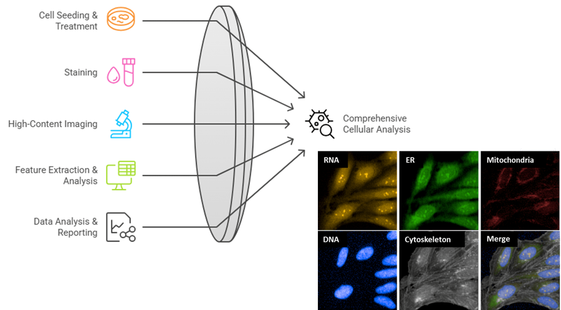
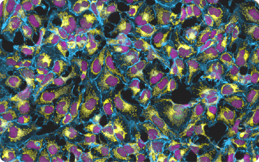
Images of U2OS cells stained with six fluorescent dyes: nucleic acids (yellow LUT), endoplasmic reticulum (green LUT), mitochondria (red LUT), DNA (blue LUT), and cytoskeleton (gray LUT). The merged image shows the overlay of all fluorescent channels.
Why choose Axxam for cell painting solutions
- Proven, reliable workflows
- Tailored solutions with full flexibility
- Fast turnaround with scalable capacity
- Stringent quality control
- Clear, consistent communication
- Seamless data analysis & expert support
- Access to in-house, commercial, or custom libraries
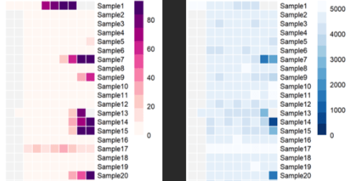
Cell painting profiling showing phenotypic induction metric (left side) and related toxic effect (right side) for each sample.
Only three steps to reach your lead candidates:
- Contact us with your biological questions and/or compound set
- We design an experimental plan with timeline & cost
- You approve → we deliver data & results