

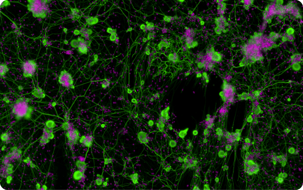
Neurons (rat DRG cells) and neurites stained with β-III Tubulin (Green LUT) in co-culture with glial cells (Pink LUT). 20X Magnification
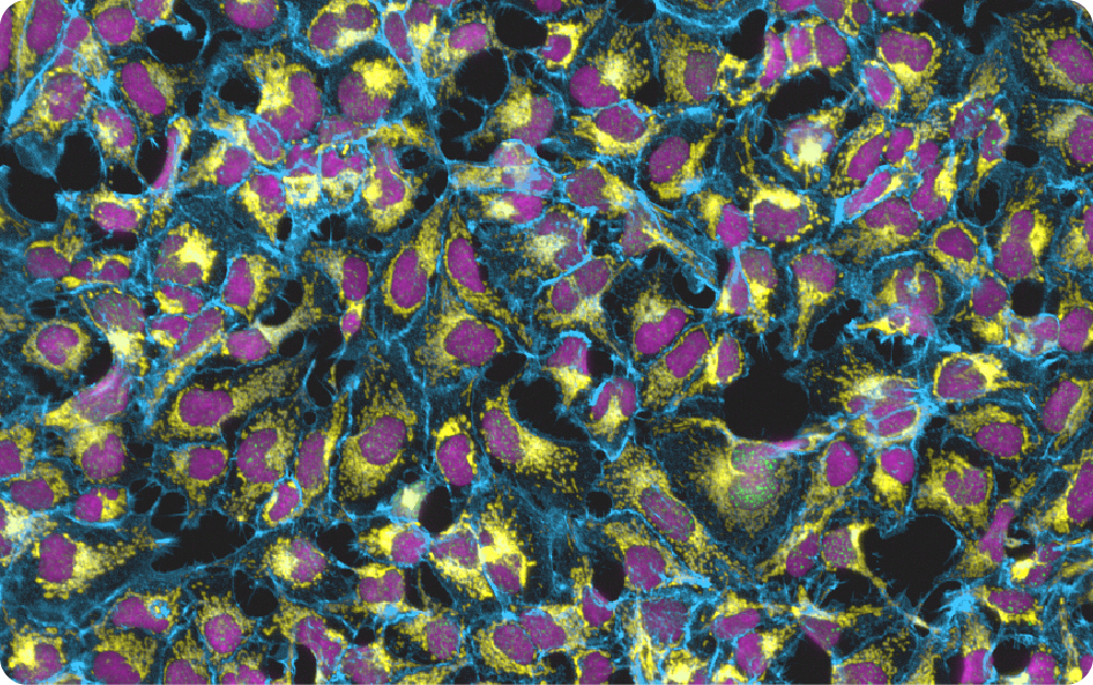
CellPainting of HeK293 cells treated with a DNA damaging agent (Etoposide). Mitochondria are stained using an anti TOM20 antibody (yellow LUT). The membrane is stained with phalloidin (blue LUT). Nuclei are labelled with DAPI (pink LUT) and γH2AX DNA damage foci are represented in green LUT. 20X Magnification
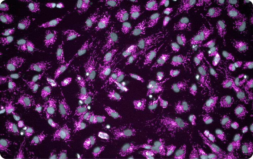
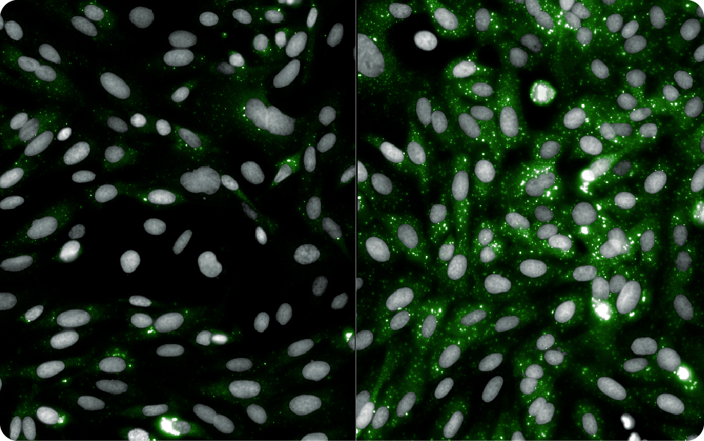
HeK293 stained with a dye labelling mitochondrion (pink LUT) and nuclei (gray LUT).
Bimolecular condensates formation by light stimulation in U2OS cancer cells. 20X Magnification
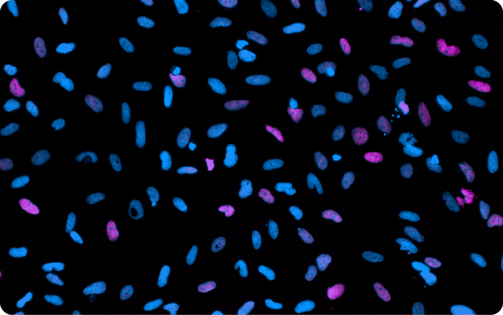
Fraction of nuclei of Human Osteosarcoma SJSA cells showing high level of Dna damage (pink Lut) upon toxic agent treatment
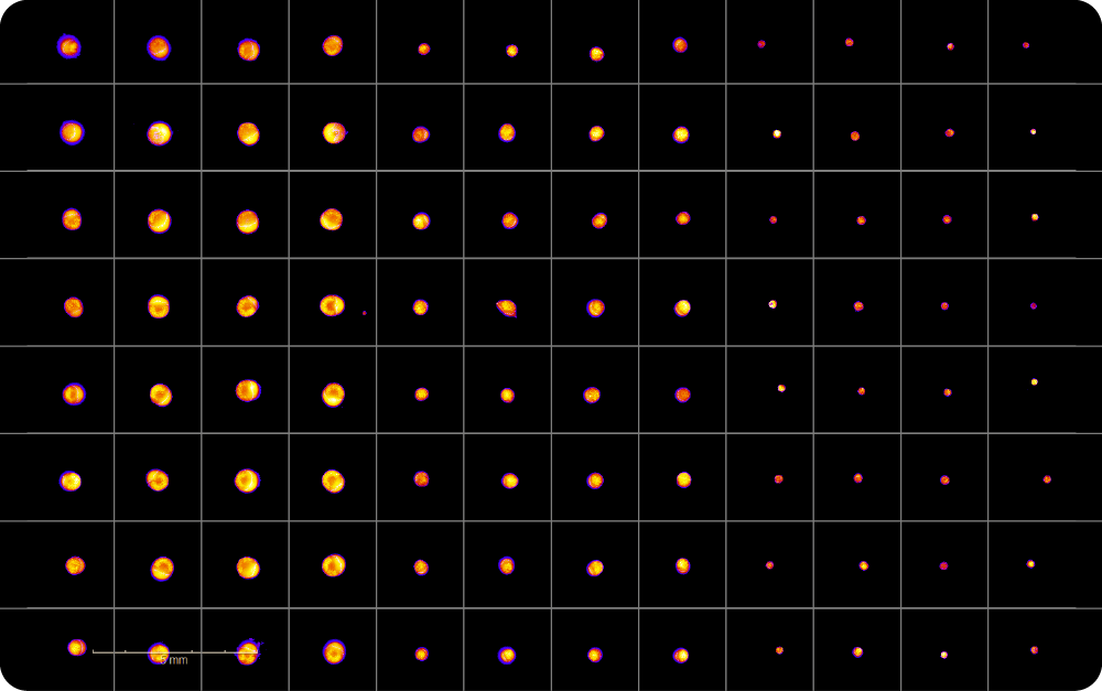
Human Osteosarcoma SJSA cells spheroids of controlled size in 96-well format. 10X Magnification